As one of the controls on the ability to generate wave replicas used sodium chloride (crystalline); sodium chloride (1M aqueous solution); tartaric acid crystals; the racemic form of tartaric acid (1M aqueous solution); air dry starch; glycine Crystal; calciferol air dry; tocopherol air dry; chlorophyll air dry; water bidistillirovannaâ; Interferon in combination with Bacillus subtilis, air dry. None of the drugs failed replicas.

 [clear]
One of the modifications of the experiment, shown in Figure 5. (the old DNA sample has been replaced by the fresh). Frames 3 and film 4. On the 4-m frame visible diode "replica links», go to the right. Typical appearance of perforation and replicas "blown highlights" near their. Frames 11 and 12. From the 4th to 11st frame replica dune no diodes, but on a 12-meter shot appear again.
[clear]
One of the modifications of the experiment, shown in Figure 5. (the old DNA sample has been replaced by the fresh). Frames 3 and film 4. On the 4-m frame visible diode "replica links», go to the right. Typical appearance of perforation and replicas "blown highlights" near their. Frames 11 and 12. From the 4th to 11st frame replica dune no diodes, but on a 12-meter shot appear again.

 [clear]
Frames 13 and 14. The 13-metre replica diodes visible dune with a characteristic into a no-go zone mežkadrovogo space. The 14-metre replica frame to disappear. Frames 23 and 24. From 14 to 22 replicas again disappear, but weakly manifested at 23 and 34 frames.
[clear]
Frames 13 and 14. The 13-metre replica diodes visible dune with a characteristic into a no-go zone mežkadrovogo space. The 14-metre replica frame to disappear. Frames 23 and 24. From 14 to 22 replicas again disappear, but weakly manifested at 23 and 34 frames.
Methods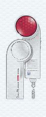
Rice. 7. Matrix with red and infrared diodes (Dune m apparatus "or" DûnaT ". http://argonet.ru/nar_lechebn_duna.htm). It contains 37 diodes, one red - 21 ( = 650nm), IR - 16 ( = 920nm).
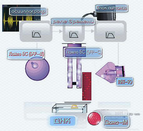
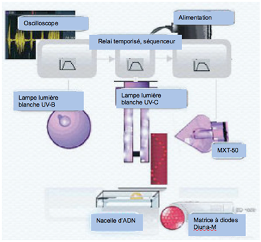
We used two circuit experiments. First, see the (Fig. 8), the second is quite simple and is visible directly on the photo (Rice. 5(in)). The schema also provides the [18].
The second version of the methods to obtain and visualize the wave display replicative DNA (Rice. 5(in)) is the following. Air dry the preparation of DNA, 100mg, placed in the clear in the aluminum foil holder. With an interval of 2-3 seconds. include lamp REF (UV-B), Compact electronic lamp CEST26E27 Black (UV-C) and "Dune". After 5 min. start photographing. In this version of the register and DNA replica objects, subject strictly to the right. At mechanical influence on drug distribution vector DNA replica changes its direction at the diametrically opposite, that is to the left. Then after 5-8 seconds. After mechanical impact, in spite of the fact, that all equipment, initiates the replica, remains enabled, replica disappear (or not committed the type of film).
Rice. 8
Rice. 8